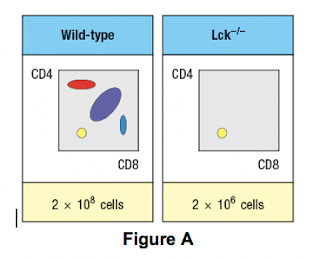
a) What is the explanation for the altered number and subsets of thymocytes in Lck-/- mice? (1 point)
Due to the defect observed in the germline Lck-deficient mice, it is not possible to use these mice to examine any potential role for this T-cell receptor signaling protein at later stages of thymocyte development. To circumvent this problem, conditional Lck-deficient mice are generated by crossing mice with a homozygous ‘floxed’ allele of Lck (Lckfl/fl) to mice that express the cre recombinase in CD4+CD8+ double-positive thymocytes (i.e., CD4-cre). When cre is expressed at the double-positive stage, the Lck gene is deleted, and thymocytes become Lck-deficient at that time. The thymocyte profile of these Lckfl/fl x CD4-cre mice is shown in the figure below.
b) What is the explanation for the altered thymocyte profile seen in Lckfl/fl x CD4-cre mice? (1 point)
To further assess the impact of altered T-cell receptor signaling on thymocyte development, another line of mice is generated that express a super-active version of the Lck kinase (called ‘Lck-super’) under control of the CD4 promoter. This super-active Lck is expressed starting at the double-positive stage in the thymus. Lck-super is activated by T-cell receptor signaling, just like wild-type Lck, but when activated has an approximately tenfold increased kinase activity. Surprisingly, mice expressing Lck-super do not develop increased numbers of mature T cells, but instead, show the following (as seen in Figure C, below).
c) What is the most likely explanation for the altered profile of thymocytes seen in the Lck-super mice? (1 point)
Struck by the findings in the Lck-super mice, an investigator performs one further experiment. This researcher clones the rearranged T-cell receptor a and b chain genes from two CD4 single-positive thymocytes: one that is isolated from a WT thymus, and the second, isolated from an Lck-super thymus. Each T-cell receptor a:b pair is used to generate a T-cell receptor transgenic line, so that nearly 100% of all double-positive thymocytes in each transgenic line express only the transgenic T-cell receptor. The two T cell receptor transgenics are named based on which mouse line the receptor chains were originally isolated from. The one from the wild-type line is known as TCR-tgwt, and the one originally isolated from the Lck-super line is known as TCR-tgsuper. In each case, thymocytes from the T-cell receptor transgenic lines are analyzed on a wild-type background, or after crossing to the Lck-super line. The results are shown in figure D, below.
d) Explain the results observed in this experiment.
2. The late 1990s saw a flurry of research identifying various TLRs in mammals, and determining their ligands.
a. What is the ligand for TLR4?
It became clear that TLR activation in macrophages could trigger the production of inflammatory molecules including IL-1, TNF, and PGE2. It was not clear, however, whether TLR signaling could activate direct killing mechanisms within macrophages themselves. The next few questions concern the analysis of this data from a paper published in 2001 addressing the role of TLR2 in immune defense against Mycobacterium tuberculosis (Thoma-Uszynski et al Science 291:1544).
b. What mycobacterial ligand is recognized by TLR2? (see text table 5-4) TLR2 TLR2- 4
c. What does it mean that the macrophages were infected with M. tuberculosis at an MOI of 5?
Figure 1. Thioglycollate-elicited macrophages from control (wt) and gene-deleted (TLR2-/-) mice were infected with M tuberculosis at an MOI of 5. 4 hours later, they were treated by the addition of the 19-kD lipoprotein (1 μg/ml) or LPS (0.1 μg/ml), 48 hours later, supernatants were assayed for NO production (Griess reaction) and cells were lysed (0.05% Tween) for plate counts of bacterial load. The figure shows the average of triplicate experiments +- SEM.
d. The 19 kD ligand used in this experiment is a cell-wall-associated lipoprotein from Mycobacterium tuberculosis that is known to activate innate immune mechanisms. The left-most panel shows that both LPS and the 19kD lipoprotein are capable of activating which innate immune mechanism? What enzyme mediates this mechanism?
e. How does the Griess reaction work (you can peek ahead to Lab 5 to see)?
3.


Post a Comment